Gulf Perio and Implants provides a variety of surgical services The practice is conservative in its treatment recommendations and often, nonsurgical solutions can be found. However, when there is no alternative and surgery is the best solution, Gulf Perio and Implants employ scientifically based procedures with state-of-the-art equipment. Beyond that, we adhere to sterilization protocols that exceed even OSHA requirements.
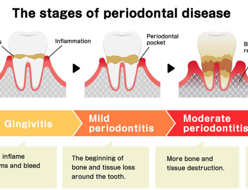
Osseous surgery, or flap surgery, is usually performed when a pocket around a tooth has not responded to other dental treatments. During the evaluation appointment, measurements are made around each tooth, giving a map of the pocket depth of each tooth. Pockets that are too deep for the patient to effectively clean, will have to be treated by a periodontist, resulting in periodontal surgery, also called osseous surgery. The goal to osseous surgery is to definitively eliminate the bacteria and infected tissue below the gum line.
In the past, gum disease was treated by eliminating the pockets by trimming away the infected gum tissue and by re-contouring the uneven bone tissue. Although this is still an effective way of treating gum disease, new and more sophisticated procedures are used routinely today. The problem with osseous resective surgery was that it often left the patient looking “long in the tooth.”
When we are consulting a patient about extracting a tooth or teeth, we like to prepare the extraction site with a bone graft. Whether the patient decides to obtain an implant or not, we like to preserve the extraction site to maintain the health of the bone. This procedure is called Ridge Preservation bone graft. The prime objective of ridge preservation is to diminish or completely eliminate the necessity of more invasive augmentation procedures in the future.
We use a revolutionary bone augmentation material called Foundation® for bone grafts and after teeth extractions.
Collagen-based, it provides support for implants, bridges, and dentures. This product stimulates bone growth and implants may be placed as early as 8-12 weeks.
Foundation
What is Foundation® and what does it do?
Foundation® is a collagen-based bone filling augmentation material indicated for use after extractions. It is not a bone substitute; rather, it stimulates growth of the patient’s own bone at an accelerated pace.
Alveolar Ridge Augmentation
Over a period of time, the jaw bone associated with missing teeth atrophies or is reabsorbed. This often leaves a condition in which there is poor quality and quantity of bone suitable for placement of an attractive bridge or dental implants. In these situations, most patients are not candidates for placement of dental implants unless the defect can be repaired.
We now have the ability to grow bone where needed with grafting techniques. This not only gives us the opportunity to place implants of proper length and width, it also gives us a chance to restore the esthetic appearance and function .
Some of these barriers are bioabsorbable and some require removal. Other regenerative procedures involve the use of bioactive gels that induce the formation of new support for the teeth.
Gum recession is one of the most noticeable results of periodontal disease. Gum recession is the movement of the gum line down the root of the tooth. When recession of the gum line occurs, the body loses a natural defense against both bacterial penetration and trauma. When gum recession is a problem, using grafting techniques is an option.
In addition, gum recession often results in root sensitivity to hot and cold foods as well as an unsightly appearance to the gum and tooth. Also, gum recession, when significant, can predispose to worsening recession and expose the root surface, which is softer than enamel, leading to root decay and root gouging.
Free Gingival Grafts
Free gingival grafts are designed to solve these problems. A thin piece of tissue is taken from the roof of the mouth. The gum graft may be placed in such a way as to cover the exposed portion of the root.
In cases with multiple areas of recession, we use Symbios PerioDerm which is an acellular dermal allograft designed to replace damaged or inadequate tissue in the repair, reinforcement or supplemental support of soft tissue defects. This procedure allows us to treat multiple areas of recession without creating a large area of palatal tissue involvement.
We are conservative in recommending this procedure but when indicated, it can result in a stable, healthy band of attached tissue around the tooth.
There are times when a two stage gingival graft are required. After allowing the gum graft to mature, a second procedure may be required.This procedure is called a Pedical Soft Tissue Graft.
Pedicle Soft Tissue Graft
The pedicle graft method does not take tissue from the roof of the mouth, instead it is gently moved over from adjacent areas, to provide a stable band of attached gingiva around the tooth. The results are excellent because the tissue that is moved includes blood vessels that are left in place.
Esthetic Crown Lengthening
A variation of crown lengthening surgery falls under the cosmetic category and is indicated when the front teeth are too short or uneven in length. Sometimes referred to as a “smile lift,” procedures are available that significantly enhance a patient’s smile and eliminate the habit of suppressing a smile or covering it with a hand!
These procedures don’t necessarily address a disease process, but they are aimed at enhancing a patient’s pride, self-esteem and quality of life by improving the esthetics of their smile:
Short teeth and upper front teeth with an uneven gum line can be exposed, restored and made to look proportionate and symmetrical
Exposed, sensitive and unsightly root surfaces can often be covered with gum grafting procedures
Concavities in the gum where teeth were previously lost can often be “plumped” to allow for a more even and pleasing appearance.
Some medications (e.g. blood pressure pills and dilantin) can cause the growth of excess gum.
It is relatively straight forward to remove that tissue and reestablish normal contours. Learn about other periodontal plastic surgical procedures.
Functional Crown Lengthening
Sometimes functional crown lengthening will be needed when decay occurs below the gum line or when a tooth fractures. It may be impossible for the General Dentist to repair the tooth without assistance from a Periodontist. In cases like this, it might be necessary to remove a small amount of bone and gum to expose more of the tooth, and give your dentist the necessary access to restore the tooth. However, simply trimming the gum line is not sufficient, instead the dentist will need to reshape the gum line and also the bone to allow adequate room for the new restoration. This minor surgical procedure allows many teeth to be saved that were previously extracted because they were deemed unrestorable.
An overly short frenum below the tongue restricts tongue movement and affects speech.
Shortened frenae on the inside of the lips pull on the gum tissue. This can cause the gum to recede away from the teeth or gaps between the teeth to develop. Frenectomies are safe, simple procedures that dramatically improve comfort and freedom of motion in your mouth.
A frenectomy is sometimes needed for dentures to fit properly, or to maintain the results of some orthodontic treatments.
A fiberotomy is a procedure to release these fibers. Fiberotomies are commonly performed toward the end of orthodontic treatment. Your orthodontist and periodontist will carefully determine which teeth are subject to repositioning.
During the procedure, your periodontist moves around the full circumference of each of these teeth, gently detaching the fibers from the gum tissue. Fiberotomies are non-invasive, predictable procedures that greatly help to preserve the results of straightening.


